Doppler color flow Veterinary ultrasound
CAD 4219.80
Overview
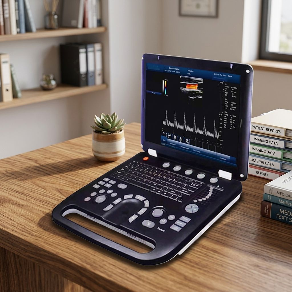
The Doppler color flow Veterinary ultrasound is designed for veterinary diagnostic imaging. B, 2B, 4B, M, B/M, B/C, B/D, B/C/D, B/CFM/D, PDI Color, Dual Color, Simultaneous 2D/Color Compound, PW, Duplex/Triplex, CFM, CDE, PD, Directional PD, CD Documented specifications include complex model imaging of IMT auto measurements, duplex & triplex synchronous display of Directional Power Doppler, usb of Flash Drive, color depth of 24bit, (2.0mhz to 10.0mhz) of Central 7.5 MHz, and (2.0mhz to 5.5mhz) of Pitch. Additional practical features include imaging compatibility with startup Time (Max):Avg. < 90 seconds, adjustable (15 steps) with beam-former Frequency Range, and imaging compatibility with tissue Harmonic Imaging Tech.
Key features
Imaging compatibility — Tissue Special Image Imaging compatibility — Panoramic Imaging Tech Imaging compatibility — Trapezoidal imaging Complex Model Imaging — IMT auto measurements USB — Flash Drive Brightness — 230 cd/m2
Technical specifications
Imaging Modes: B, 2B, 4B, M, B/M, B/C, B/D, B/C/D, B/CFM/D, PDI Color, Dual Color, Simultaneous 2D/Color Compound, PW, Duplex/Triplex, CFM, CDE, PD, Directional PD, CD Probe: Convex Array Probe Linear Array Probe: Intra-Cavity Probe Frequency: Central 3.5 MHz (2.0mhz to 10.0mhz): Central 7.5 MHz (6.0mhz to 10.0mhz): Central 6.5 MHz (5.0mhz to 9.0mhz): Central 4.0 MHz (2.0mhz to 5.5mhz): Pitch 0.516mm: 0.352mm 0.216mm: Radius 60mm: N/A 10 Mm: Elements Imaging Performance: Startup Time (Max):Avg. < 90 seconds Preset Switching Time: Avg. < 1 second Storage Time (image to Disk): Avg. < 0.5 second Hardware Specification: Size (Diagonal) Contrast Ratio: 800:1 Resolution: 1024×768 pixels Brightness: 230 cd/m2 Color Depth: 24bit Grey Levels: 256 An/zoom: Real-Time Image Zoom, Zoom Range: 100%~400%, Up/Down/Left/Right Inversion General Performance: Digital Broadband 12288 Channels: Beam-former Re-programmable: Transmit Voltage Adjustable (15 Steps): Beam-former Frequency Range 1~40 Mhz: CF+B mode in one screen Complex Model Imaging: IMT auto measurements Virtual Convex Array: Trapezoidal imaging Duplex & Triplex Synchronous Display: Directional Power Doppler Imaging Parameters Preset: Tissue Special Image Pw Auto Trace: Update in line Technology: Panoramic Imaging Tech All-digital Signal Processing Tech: Multi-beam formation Tech Speckle Reduction Tech: Tissue Harmonic Imaging Tech Dynamic Tissue Optimization Tech: DICOM Connectivity Dicom3.0 Compliant: 3D Software Built-in 3d Software: Image Storage Storage Format: PNG, AVI, BMP, JPEG, DICOM USB: Flash Drive
Dimensions
Rotate Angle: ± 90°